JEE Advanced (IIT Entrance) · Chemistry (JEE & NEET)
Biomolecules
Structure and functions of carbohydrates, proteins, amino acids, enzymes, vitamins, nucleic acids and hormones.
Eleven concepts covering carbohydrates (roles and classification, glucose preparation and cyclic forms, reducing sugars, di- and polysaccharides), amino acids and proteins (zwitterions and essentials, peptide bonds and structure levels including fibrous versus globular, denaturation), nucleic acids (DNA versus RNA, base pairing), vitamins (fat- and water-soluble sources and deficiencies), and enzymes as specific catalysts. Most marks are classifications — reducing or not, fibrous or globular, DNA or RNA, fat-soluble or water-soluble — with one Chargaff arithmetic check from hydrogen-bond counts.
- JEE Advanced (IIT Entrance)
- Easy level
- 11 concepts
- 5 practice questions
1Carbohydrates: roles and classification
Carbohydrates are polyhydroxy aldehydes or ketones, or compounds that hydrolyse to them. They are the chief source of energy in living organisms (glucose oxidation), the storage form of that energy (starch in plants, glycogen in animals), and the structural scaffolding of plant cell walls (cellulose).
Hydrolysis depth splits them into monosaccharides (cannot be hydrolysed further), oligosaccharides (hydrolyse to two–ten monosaccharide units; disaccharides are the usual case), and polysaccharides (hydrolyse to many units). An independent cut names a monosaccharide by carbonyl type and carbon count: an aldose carries an aldehyde, a ketose a ketone; triose, tetrose, pentose, hexose finish the name. Glucose is an aldohexose (\mathrm{C_6H_{12}O_6}); fructose is a ketohexose with the same molecular formula.
Figure. Hydrolysis classification of carbohydrates: monosaccharides give no further sugar on hydrolysis; oligosaccharides give two–ten monosaccharide units (disaccharides are the usual exam cases); polysaccharides give many. Aldose/ketose × carbon count is a second cut on monosaccharides — carried by this concept's class table, not by this tree.
How it works
- Ask what it doesEnergy now (glucose), energy stored (starch, glycogen), or structure (cellulose) — three roles, not three formulas.
- Ask if it hydrolysesNo further hydrolysis → mono. Two–ten units on hydrolysis → oligo (disaccharides dominate). Many units → poly.
- Name a monosaccharideCarbonyl first (aldose or ketose), then carbon count. Aldo + hexose → aldohexose. Same \mathrm{C_6H_{12}O_6} does not decide glucose versus fructose.
| Sugar | Carbonyl | Carbons | Class |
|---|---|---|---|
| Glucose | Aldehyde | 6 | Aldohexose |
| Fructose | Ketone | 6 | Ketohexose |
| Ribose | Aldehyde | 5 | Aldopentose |
| Glyceraldehyde | Aldehyde | 3 | Aldotriose |
Fructose is classified as a
- aldohexose, because it has six carbons and reduces Tollens' reagent
- ketohexose — six carbons and a ketone carbonyl
- ketopentose, because a five-membered ring is common
Fructose has six carbons and a ketone carbonyl, so it is a ketohexose. Ring size (furanose versus pyranose) is a separate fact and does not rename the open-chain class.
2Glucose preparation and cyclic forms
Glucose is obtained by acid hydrolysis of sucrose: \mathrm{C_{12}H_{22}O_{11} + H_2O \xrightarrow{H^+} C_6H_{12}O_6 + C_6H_{12}O_6}, giving one molecule each of glucose and fructose. It is also obtained from starch or cellulose: (\mathrm{C_6H_{10}O_5})_n + n\,\mathrm{H_2O} \xrightarrow{\text{dil.\,H}_2\mathrm{SO}_4,\,393\,\mathrm{K},\,\text{pressure}} n\,\mathrm{C_6H_{12}O_6}.
In water the open-chain aldehyde of glucose closes to a six-membered hemiacetal ring (pyranose). The new chiral centre at C-1 is the anomeric carbon: α and β anomers differ only in the configuration there. Fructose closes to a five-membered hemiketal (furanose) or a six-membered pyranose; sucrose uses the β-D-fructofuranose form.
Figure. Acid hydrolysis of sucrose gives glucose + fructose. Each open chain closes to a hemiacetal/hemiketal ring — pyranose (six) or furanose (five) — with α/β anomers at the anomeric carbon. Ring names are the exam ask without Haworth art.
How it works
- From sucroseDilute acid hydrolyses the glycosidic bond → equimolar glucose + fructose. Invert sugar is that mixture.
- From starch or cellulose(\mathrm{C_6H_{10}O_5})_n + n\,\mathrm{H_2O} \rightarrow n\,\mathrm{C_6H_{12}O_6} under dilute \mathrm{H_2SO_4} at 393 K and pressure.
- Close the chainGlucose: OH of C-5 attacks C-1 → pyranose; α/β at C-1. Fructose: OH of C-5 (or C-6) attacks C-2 → furanose or pyranose.
| Source / form | Conditions or ring | Product / note |
|---|---|---|
| Sucrose | Dilute acid, warm | Glucose + fructose (1:1) |
| Starch / cellulose | Dil. H₂SO₄, 393 K, pressure | n glucose |
| D-Glucose cyclic | Pyranose (6-membered) | α / β anomers at C-1 |
| D-Fructose cyclic | Furanose or pyranose | Anomeric carbon is C-2 |
Acid hydrolysis of sucrose under the usual laboratory conditions yields
- only glucose, because fructose is destroyed by acid
- equimolar glucose and fructose
- only fructose, because glucose stays bound
Sucrose is a glucose–fructose disaccharide; acid cleavage of its glycosidic bond frees one molecule of each. Starch hydrolysis, not sucrose, is the route that yields glucose alone.
3Reducing and non-reducing sugars
A reducing sugar has a free anomeric carbon — one that can open to expose an aldehyde (or an α-hydroxy ketone that tautomerises to one). That free carbonyl reduces Tollens' reagent (silver mirror) and Fehling's solution (Cu₂O precipitate). Glucose, fructose, maltose and lactose are reducing.
Sucrose is the standard non-reducing exception: its glycosidic bond joins the anomeric carbon of glucose (C-1) to the anomeric carbon of fructose (C-2), so neither end can open. If a disaccharide's linkage leaves any anomeric carbon free, the sugar is reducing.
Figure. Decision tree for reducing status: free anomeric carbon → reducing (glucose, maltose, lactose, fructose); both anomerics locked → non-reducing (sucrose). The glycosidic bond itself is not drawn — only the yes/no criterion.
How it works
- Look at the anomeric carbonFree hemiacetal/hemiketal → can open → reducing. Both anomerics tied in a full acetal → non-reducing.
- MonosaccharidesGlucose and fructose are reducing even though fructose is a ketose — it tautomerises under the test conditions.
- DisaccharidesMaltose and lactose each leave one anomeric free. Sucrose locks both — the exam's favourite trap.
| Sugar | Free anomeric? | Tollens' / Fehling's | Class |
|---|---|---|---|
| Glucose | Yes | Positive | Reducing |
| Fructose | Yes (after tautomerism) | Positive | Reducing |
| Maltose | One free | Positive | Reducing |
| Lactose | One free | Positive | Reducing |
| Sucrose | Neither free | Negative | Non-reducing |
Sucrose does not reduce Tollens' reagent because
- it contains no oxygen atoms that could be oxidised
- both anomeric carbons are locked in the glycosidic bond
- it is a polysaccharide, and only monosaccharides reduce
Sucrose joins C-1 of glucose to C-2 of fructose, so neither anomeric carbon can open to a free carbonyl. Maltose and lactose each leave one anomeric free and do reduce.
4Di- and polysaccharides
Three disaccharides carry most of the marks. Maltose is two α-D-glucose units joined by an α-1,4-glycosidic linkage and is reducing (one anomeric free). Lactose is β-D-galactose joined to β-D-glucose by a β-1,4 linkage and is likewise reducing. Sucrose joins C-1 of α-D-glucose to C-2 of β-D-fructose, locks both anomerics, and is non-reducing.
Starch is the plant storage polysaccharide: amylose is an unbranched α-1,4 chain of D-glucose; amylopectin is branched, with α-1,4 links in the chains and α-1,6 links at the branch points. Glycogen is the animal storage form — like amylopectin but more highly branched. Cellulose is a structural β-1,4 polymer of D-glucose; humans lack the enzyme that cleaves that β linkage.
Figure. Disaccharide identity is the linkage: maltose α-1,4 and lactose β-1,4 stay reducing; sucrose ties both anomerics (α-1,β-2) and is non-reducing. Polysaccharides add branching (α-1,6) on top of α-1,4 chains.
How it works
- Name the units and the linkMaltose: Glc–α-1,4–Glc. Lactose: Gal–β-1,4–Glc. Sucrose: Glc–α-1,β-2–Fru.
- Ask which anomeric is freeOne free → reducing (maltose, lactose). Both tied → non-reducing (sucrose).
- Storage versus structureStarch (amylose + amylopectin) and glycogen store glucose with α links. Cellulose builds walls with β-1,4 links.
| Name | Units / monomer | Linkage | Note |
|---|---|---|---|
| Maltose | α-D-Glc + α-D-Glc | α-1,4 | Reducing |
| Lactose | β-D-Gal + β-D-Glc | β-1,4 | Reducing |
| Sucrose | α-D-Glc + β-D-Fru | α-1,β-2 | Non-reducing |
| Amylose | α-D-Glc | α-1,4 only | Linear; plant storage |
| Amylopectin | α-D-Glc | α-1,4 + α-1,6 | Branched; plant storage |
| Glycogen | α-D-Glc | α-1,4 + α-1,6 | More branched; animal storage |
| Cellulose | β-D-Glc | β-1,4 only | Linear; plant structure |
Amylopectin differs from amylose mainly in that amylopectin
- is built from β-D-glucose with β-1,4 links only
- has α-1,6 branch points in addition to α-1,4 chain links
- is the animal storage polysaccharide, not a plant one
Both amylose and amylopectin are plant starch fractions of α-D-glucose. Amylose is unbranched α-1,4; amylopectin adds α-1,6 branches. β-1,4 is cellulose; the animal store is glycogen.
5Amino acids and zwitterions
α-Amino acids carry an amino group and a carboxyl group on the same (α) carbon. They are amphoteric: the –COOH can donate a proton and the –NH₂ can accept one. In the solid and in aqueous solution near neutrality they exist as zwitterions, ^{+}\mathrm{H_3N{-}CHR{-}COO^-}, not as the uncharged \mathrm{H_2N{-}CHR{-}COOH} form.
The isoelectric point (pI) is the pH at which the net charge is zero — the zwitterion dominates and the amino acid does not migrate in an electric field. Side chains sort the twenty proteinogenic acids into acidic (Asp, Glu — an extra carboxyl), basic (Lys, Arg, His — an extra amino-like nitrogen), and neutral (equal acidic and basic groups on the α-framework).
Essential amino acids cannot be synthesised by humans at a rate that meets need and must come from the diet. NCERT lists ten essentials: Val, Leu, Ile, Phe, Thr, Trp, Met, Lys, Arg and His. The rest are non-essential — the body can make them — even though Arg and His are sometimes called semi-essential outside the NCERT list.
Figure. The zwitterion ⁺H₃N–CHR–COO⁻ dominates near the isoelectric point pI, where net charge is zero. Lower pH protonates the carboxylate (+1); higher pH deprotonates the ammonium (−1).
How it works
- Two ionisable groups–COOH is acidic; –NH₃⁺ is acidic too once protonated. An acidic or basic side chain may add a third ionisable group and shift the pI.
- Zwitterion at pIAt the isoelectric pH the average charge is zero: ^{+}\mathrm{H_3N{-}CHR{-}COO^-}. Below pI the cationic form dominates; above pI the anionic form does.
- Side-chain classAcidic = Asp, Glu. Basic = Lys, Arg, His. Neutral = the others (Gly, Ala, Ser, …). Class follows extra –COOH versus extra –NH₂ on the side chain.
- Essential vs non-essentialEssential means diet-required. NCERT's ten: Val, Leu, Ile, Phe, Thr, Trp, Met, Lys, Arg, His. Non-essential means the body can synthesise them.
For a simple (neutral side-chain) α-amino acid relative to its own pI.
| Relative pH | Dominant form | Net charge |
|---|---|---|
| pH ≪ pI | ⁺H₃N–CHR–COOH | Positive |
| pH = pI | ⁺H₃N–CHR–COO⁻ (zwitterion) | Zero |
| pH ≫ pI | H₂N–CHR–COO⁻ | Negative |
According to the NCERT Class 12 list, which set is entirely essential amino acids?
- Glycine, alanine and serine
- Valine, lysine, arginine and histidine
- Aspartic acid, glutamic acid and tyrosine
NCERT's ten essentials are Val, Leu, Ile, Phe, Thr, Trp, Met, Lys, Arg and His — so Val, Lys, Arg and His are all on that list. Gly/Ala/Ser are non-essential; Asp/Glu/Tyr are likewise non-essential (Asp and Glu are the acidic pair).
6Peptide bonds and protein structure levels
Proteins are polymers of α-amino acids joined by peptide (amide) bonds, written -\mathrm{CO{-}NH}-. The carboxyl carbon of one residue links to the amino nitrogen of the next, releasing water. The sequence of residues is the primary structure — and it alone determines everything that folds later.
Secondary structure is local backbone folding: the α-helix and the β-pleated sheet, both held by hydrogen bonds between backbone \mathrm{C{=}O} and \mathrm{N{-}H}. Tertiary structure is the full three-dimensional fold of one chain; quaternary structure is how multiple folded chains pack together (haemoglobin's four subunits).
Shape and solubility sort proteins into fibrous and globular. Fibrous proteins are fibre-like and generally water-insoluble — keratin (hair, nails), collagen (tendons) and myosin (muscles). Globular proteins are roughly spherical and usually water-soluble — enzymes, haemoglobin and albumin. The helix and sheet are three-dimensional objects this vocabulary cannot draw honestly.
Figure. The four structure levels as a hierarchy: sequence → local fold → one-chain 3D → multi-chain assembly. Bar and ribbon geometry of the α-helix and β-sheet are not drawn — only the naming ladder the exam uses. Fibrous versus globular is a separate shape/solubility sort, carried by the topic-level table.
How it works
- Peptide bond–COOH of residue i + –NH₂ of residue i+1 → –CO–NH– + H₂O. Planar amide; restricted rotation about the C–N bond.
- Primary → secondarySequence first (covalent peptide bonds). Then local H-bonded patterns: α-helix or β-sheet.
- Tertiary → quaternaryOne chain's full fold is tertiary (side-chain interactions). Multiple chains assembling is quaternary — absent in a single-chain protein.
- Fibrous vs globularFibrous: elongated, insoluble — keratin, collagen, myosin. Globular: compact, usually soluble — enzymes, haemoglobin, albumin.
| Level | What it names | Held by |
|---|---|---|
| Primary | Amino-acid sequence | Covalent peptide bonds |
| Secondary | α-helix, β-sheet | Backbone H-bonds |
| Tertiary | 3D fold of one chain | Side-chain interactions |
| Quaternary | Multi-chain assembly | Same, between subunits |
Keratin and collagen are examples of
- globular proteins, because they are enzymes
- fibrous proteins — fibre-like and generally water-insoluble
- quaternary structure only, because they always have four chains
NCERT classes keratin (hair/nails) and collagen (tendons) with myosin as fibrous proteins. Enzymes, haemoglobin and albumin are the globular examples. Quaternary structure is a folding level, not a fibrous/globular label.
7Protein denaturation
Denaturation is loss of the native fold: secondary, tertiary and — when present — quaternary structure are disrupted, so the protein loses its biological activity. The primary structure, the covalent sequence of peptide bonds, remains intact.
Heat, a sharp change of pH, urea, heavy-metal salts and organic solvents are the usual agents — they break the non-covalent interactions that hold the fold. Boiling egg albumin is the textbook coagulation example: the white goes from clear and soluble to opaque and solid as the chains unfold and tangle. Curdling of milk (lactic acid from bacteria) is the same idea at lower temperature.
Because activity needs the native shape, a denatured enzyme or haemoglobin cannot do its job even though every peptide bond is still there. Renaturation is sometimes possible if conditions return gently; irreversible coagulation is common once chains aggregate.
Figure. Native fold → denatured chain under heat, pH or urea. The figure names which levels vanish and which survive; it does not invent a 3D ribbon of the fold or a numerical activity–temperature curve.
How it works
- What is lostH-bonds, hydrophobic packing, ionic links and (where present) subunit contacts break. α-helix and β-sheet unroll; the chain opens.
- What survivesPrimary structure — the –CO–NH– sequence — is covalent and stays. Denaturation is not hydrolysis of the backbone.
- AgentsHeat; extreme pH; urea; heavy-metal salts; organic solvents. Each loosens the non-covalent fold.
- ConsequenceBiological activity collapses. Egg albumin coagulates on boiling; milk curdles when acid unfolds casein.
| Agent | Typical effect | NCERT-style example |
|---|---|---|
| Heat | Breaks H-bonds; unfolds chain | Boiling egg albumin |
| pH change | Alters charge; disrupts ionic links | Milk curdling (lactic acid) |
| Urea | Competes for H-bonds | Laboratory unfolding |
| Heavy-metal salts | Bind side-chain groups | Protein precipitation |
| Organic solvents | Disturb hydrophobic packing | Alcohol denaturation |
When egg albumin is boiled and coagulates, which statement is correct?
- Peptide bonds hydrolyse, so the primary sequence is destroyed
- Secondary and tertiary structure are lost; the primary sequence remains
- Only quaternary structure is affected, because albumin has four subunits
Denaturation disrupts 2°/3°/4° folds but leaves the covalent primary sequence intact. Boiling does not hydrolyse the peptide backbone; albumin's coagulation is the NCERT example of that loss of native fold and activity.
8DNA versus RNA
Nucleic acids are polymers of nucleotides. A nucleoside is a nitrogenous base linked to a pentose sugar; a nucleotide is that nucleoside plus a phosphate group. The bases split into purines — adenine (A) and guanine (G) — and pyrimidines — cytosine (C), thymine (T) in DNA, and uracil (U) in RNA.
In the chain, the phosphate joins the 3′-OH of one sugar to the 5′-OH of the next: a phosphodiester backbone with a free 5′ end and a free 3′ end. DNA uses 2-deoxyribose and thymine; RNA uses ribose and uracil. DNA has thymine and deoxyribose; RNA has uracil and ribose — swapping either marker is the classic trap.
Figure. A nucleotide is three parts: nitrogenous base, pentose sugar and phosphate. Drop the phosphate and what remains is a nucleoside. Purine versus pyrimidine, 2-deoxyribose versus ribose, and thymine versus uracil then decide DNA versus RNA — that split sits in this concept's comparison table. The helix itself is not drawn.
How it works
- Name the unitNucleoside = base + sugar. Nucleotide = base + sugar + phosphate.
- Sort the basesPurines: A, G (two rings). Pyrimidines: C, T, U (one ring). DNA uses T; RNA uses U.
- Link the chain3′–5′ phosphodiester bonds join successive sugars; the strand direction is written 5′ → 3′.
- Mark DNA vs RNADNA: 2-deoxyribose + T, usually double helix. RNA: ribose + U, usually single-stranded.
| Feature | DNA | RNA |
|---|---|---|
| Sugar | 2-Deoxyribose | Ribose |
| Unique pyrimidine | Thymine (T) | Uracil (U) |
| Purines | A, G | A, G |
| Pyrimidines | C, T | C, U |
| Backbone | 3′–5′ phosphodiester | 3′–5′ phosphodiester |
| Usual form | Double helix (antiparallel) | Single-stranded (local folds) |
| Main role | Long-term genetic store | Transcript / adapter / catalyst |
A nucleic acid that contains uracil and ribose is
- DNA, because uracil pairs with adenine in the double helix
- RNA — uracil and ribose are the RNA markers
- either DNA or RNA; the bases alone do not decide
Uracil and ribose identify RNA. DNA uses thymine and 2-deoxyribose instead.
9Base pairing and hydrogen bonds
In DNA, adenine pairs with thymine and guanine pairs with cytosine. Chargaff's rules follow at once: mole percent A equals T, and G equals C. The A–T pair is held by two hydrogen bonds; the G–C pair by three. A G–C-rich duplex therefore has more H-bonds per base pair and a higher melting temperature.
That is why a stem that gives one base percent is enough to recover all four, and why stability arguments point at the triple-bonded G–C pair rather than at the length of the chain alone.
Figure. Base composition for the worked example (30% A). Chargaff forces T = 30% and G = C = 20%. Annotations recall the H-bond counts: two in each A–T pair, three in each G–C pair — the reason G–C-rich DNA melts higher. Bar heights are the percents by construction.
How it works
- Pair the basesA = T (two H-bonds) and G ≡ C (three). One percent fixes its partner; the other two share what remains, equally.
- Count H-bondsEach A–T pair contributes 2; each G–C pair contributes 3. Sum over all pairs for the duplex.
- Read stabilityHigher G–C content → more triple H-bonds → higher melting temperature.
Hydrogen bonds in a DNA segment
A DNA segment has 30% adenine. Find the percent of each base. For 100 bases (50 pairs), find the total number of hydrogen bonds between strands.
- A = 30% ⇒ T = 30% (A pairs with T)A = T = 30%
- G + C = 100 − 6040%; G = C = 20%
- Pairs in 100 bases: 30 A–T + 20 G–C50 pairs
- H-bonds = 30×2 + 20×360 + 60 = 120
Pro tip. Average H-bonds per pair here is 120/50 = 2.4 — between pure A–T (2) and pure G–C (3). Raise the G–C percent and both the average and the melting temperature climb.
A double-stranded DNA has 20% guanine. The percentage of adenine is
- 20%
- 30%
- 60%
G = 20% ⇒ C = 20%. A + T = 60%, and A = T, so A = 30%.
10Fat-soluble and water-soluble vitamins
Vitamins are organic micronutrients needed in small amounts. They split by solubility: A, D, E and K are fat-soluble and can be stored in adipose tissue; the B-complex vitamins and vitamin C are water-soluble and are not stored to the same degree, so a regular dietary supply matters more.
NCERT pairs each vitamin with sources and a deficiency disease — night blindness or xerophthalmia for A, rickets for D, scurvy for C, beri-beri for B₁, pernicious anaemia for B₁₂, and longer clotting time for K. Solubility class and deficiency name are asked as a pair more often than either alone.
Figure. Two solubility classes side by side. Fat-soluble vitamins A, D, E, K can accumulate in adipose tissue; water-soluble B-complex and C do not. Sources and deficiency names sit in this concept's vitamin table — the figure only carries the split.
How it works
- Split by solubilityFat-soluble: A, D, E, K. Water-soluble: B-complex and C.
- Storage consequenceFat-soluble vitamins accumulate in adipose tissue; water-soluble ones clear more readily.
- Link source and diseaseRead the vitamin table in this concept — each row ties solubility, NCERT sources and the deficiency name.
| Vitamin | Solubility | Sources (NCERT) | Deficiency disease |
|---|---|---|---|
| A (retinol) | Fat | Fish liver oil, carrots, butter, milk | Night blindness; xerophthalmia |
| D (calciferol) | Fat | Sunlight, fish, egg yolk | Rickets; osteomalacia |
| E (tocopherol) | Fat | Wheat germ oil, sunflower oil | Fragile RBCs; muscle weakness |
| K | Fat | Green leafy vegetables | Increased clotting time |
| B₁ (thiamine) | Water | Yeast, milk, green vegetables, cereals | Beri-beri |
| B₂ (riboflavin) | Water | Milk, egg white, liver, kidney | Cheilosis; skin burning |
| B₆ (pyridoxine) | Water | Yeast, milk, egg yolk, cereals, grams | Convulsions |
| B₁₂ | Water | Meat, fish, egg, curd | Pernicious anaemia |
| C (ascorbic acid) | Water | Citrus fruits, amla, leafy vegetables | Scurvy (bleeding gums) |
Night blindness is caused by deficiency of
- vitamin C, which is water-soluble
- vitamin A, which is fat-soluble
- vitamin K, which is needed for clotting
Vitamin A deficiency → night blindness (and xerophthalmia). C is scurvy, K is longer clotting time — both real links, wrong disease for this stem.
11Enzymes as specific catalysts
Enzymes are biological catalysts, almost always proteins, that act on a narrow set of substrates at an active site. Catalysis lowers the activation energy of the reaction; the enzyme is regenerated and not consumed, so one molecule turns over many substrate molecules.
Specificity is the exam idea: the active site recognises shape and chemistry, so one enzyme does not stand in for another. Harsh heat or extreme pH can denature the fold that builds that site and kill activity — the same fragility that sets temperature and pH optima — without changing the definition of catalysis itself.
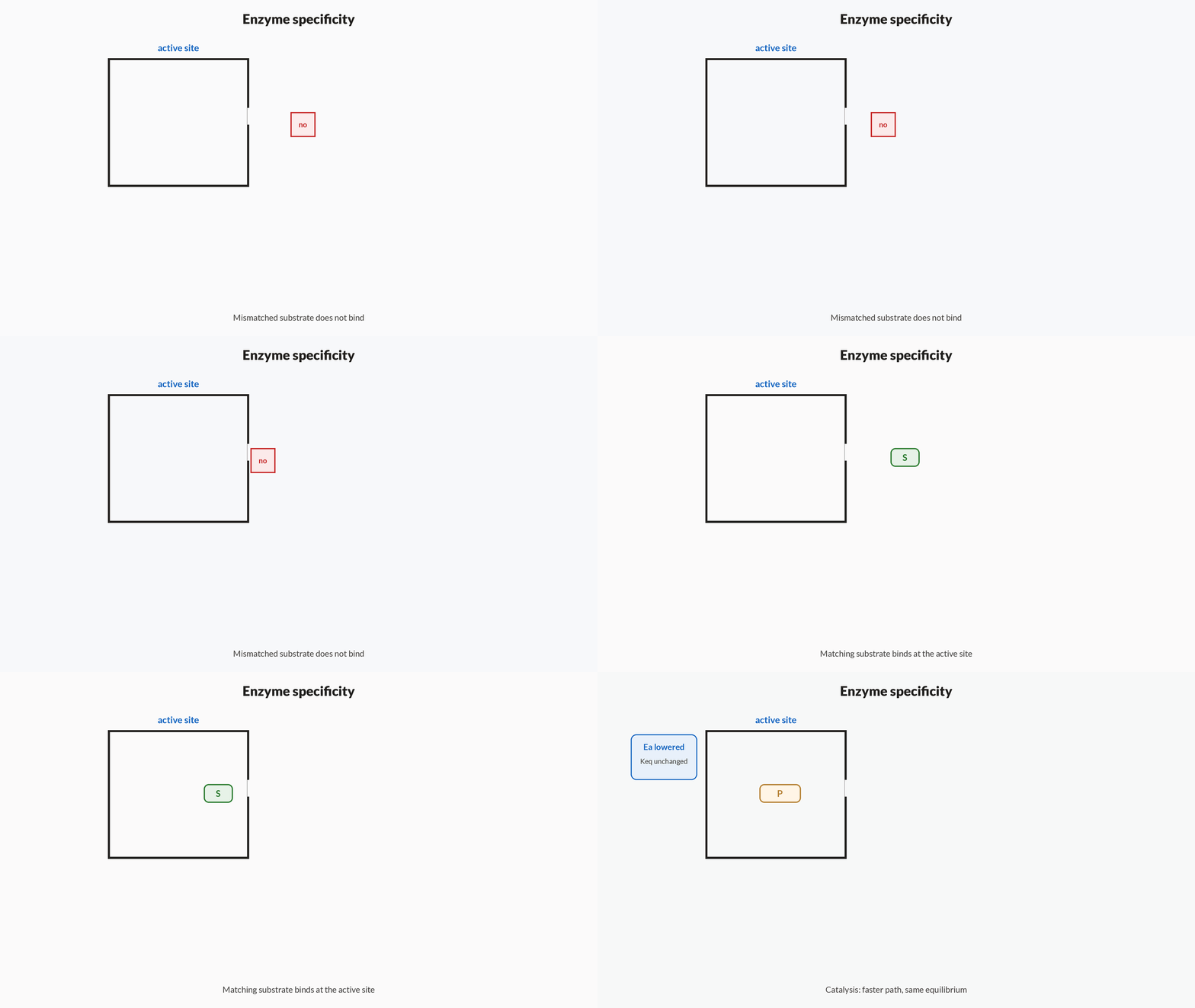
How it works
- Active siteThe folded protein creates a pocket that binds one substrate (or a tight family).
- Lower EaBinding and catalytic groups cut the activation barrier; rate rises, ΔG of the reaction is unchanged.
- RegeneratedProduct leaves; free enzyme turns over again. It is a catalyst, not a reactant.
- Fragile foldDenaturation destroys the active-site geometry, so activity falls — a brief link to protein structure, not a second concept.
Enzymes are described as highly specific because
- each enzyme is consumed after a single turnover
- each active site recognises only a narrow set of substrates
- they raise the activation energy of every reaction equally
Specificity means the active site fits a narrow substrate set. Enzymes lower activation energy and are regenerated, not consumed.
Notes
- Carbohydrates are polyhydroxy aldehydes or ketones: glucose is an aldohexose (pyranose ring) and fructose a ketohexose; sucrose is non-reducing, while maltose and lactose are reducing.
- Proteins are polymers of alpha-amino acids joined by peptide (amide) bonds, with primary, secondary (alpha-helix, beta-sheet), tertiary and quaternary levels of structure.
- Amino acids are amphoteric and exist as zwitterions at the isoelectric point (pI); essential amino acids must be obtained from the diet.
- Nucleic acids: DNA (deoxyribose; A-T, G-C) stores genetic information as a double helix, while RNA (ribose; uracil) is usually single-stranded.
- Vitamins and enzymes: fat-soluble vitamins (A, D, E, K) and water-soluble ones (B, C); enzymes are highly specific protein catalysts.
- Carbohydrates: distinguish aldoses/ketoses, reducing vs non-reducing sugars (sucrose is non-reducing because both anomeric carbons are locked in the glycosidic bond), and understand mutarotation (interconversion of alpha and beta anomers of glucose via the open-chain form, changing optical rotation to an equilibrium value).
- Glucose structure was established by key evidence: it forms a pentaacetate (five -OH), gives an oxime and adds HCN (a -CHO group), is oxidised by bromine water to gluconic acid (aldehyde) and by HNO3 to saccharic acid (both ends oxidisable), and Fischer determined the D-configuration.
- Amino acids are amphoteric, existing as zwitterions; the isoelectric point (pI) is the pH of zero net charge. Peptide bonds (amide) form between the -COOH of one and -NH2 of the next; the peptide bond has partial double-bond character (planar, restricted rotation).
- Protein structure hierarchy: primary (sequence), secondary (alpha-helix, beta-pleated sheet stabilised by H-bonds), tertiary (3D folding via disulfide, ionic, H-bonds, hydrophobic), quaternary (subunit assembly). Denaturation disrupts secondary and higher structure without breaking peptide bonds.
- Nucleic acids: nucleoside (base + sugar) vs nucleotide (base + sugar + phosphate); DNA (deoxyribose, thymine, double helix, Chargaff base pairing A=T, G=C) versus RNA (ribose, uracil, usually single-stranded). Understand the 3'-5' phosphodiester backbone.
Formulas
- Glucose molecular formula: C_6H_{12}O_6
- Peptide bond: -CO-NH- (amide linkage)
- DNA base pairing: A=T (2 H-bonds), G≡C (3 H-bonds)
- Reducing sugars give positive Tollens' and Fehling's tests (free –CHO)
- Nucleotide = base + sugar + phosphate; nucleoside = base + sugar
- \text{pI} = \dfrac{pK_{a1} + pK_{a2}}{2}\;(\text{neutral amino acid})
- \text{Glucose} \xrightarrow{Br_2/H_2O} \text{gluconic acid (C1 -CHO oxidised)}
- \text{Glucose} \xrightarrow{HNO_3} \text{saccharic acid (C1 and C6 oxidised)}
- \text{Chargaff: } [A]=[T],\;[G]=[C]\;(\text{ds-DNA})
Exam traps & shortcuts
- Sucrose is non-reducing because both anomeric carbons form the glycosidic bond; glucose, fructose, maltose and lactose are reducing.
- Deficiency links: vitamin A → night blindness, C → scurvy, D → rickets, B₁ → beri-beri, K → poor clotting.
- DNA has thymine and deoxyribose; RNA has uracil and ribose.
- Sucrose is non-reducing (glycosidic linkage between both anomeric carbons of glucose and fructose); maltose and lactose are reducing (one free anomeric carbon).
- To decide net charge of an amino acid at a given pH: below pI it is net positive, above pI net negative; at pI it is a neutral zwitterion and does not migrate in electrophoresis.
- Mild oxidation (Br2 water) touches only the aldehyde (-> monoacid); strong oxidation (HNO3) attacks both terminal carbons (-> dicarboxylic saccharic acid). Use this to infer functional groups.
Reference tables
Compact identities for night-before scanning. Concept tables carry the full decision charts.
| Identity | Statement | Watch |
|---|---|---|
| Invert sugar | Acid-hydrolysed sucrose = glucose + fructose | Equimolar mix |
| Starch → glucose | (C₆H₁₀O₅)ₙ + n H₂O; dil. H₂SO₄, 393 K, pressure | Cellulose same product |
| Sucrose link | Glc C-1 to Fru C-2 | Non-reducing |
| Cellulose link | β-1,4 D-glucose | Structural; humans lack the enzyme |
| Zwitterion | ⁺H₃N–CHR–COO⁻ at pI | Net charge zero |
| Peptide bond | –CO–NH– between residues | Primary structure is the sequence |
| Denaturation | Loses 2°/3°/4°; primary stays | Activity collapses |
| Nucleotide | base + sugar + phosphate | Nucleoside drops the phosphate |
| DNA markers | 2-deoxyribose + thymine | Do not swap with RNA |
| RNA markers | ribose + uracil | U replaces T |
| DNA base pairing | A=T (2 H-bonds), G≡C (3) | G–C-rich DNA melts higher |
| Chargaff | %A = %T and %G = %C | One percent recovers all four |
The yes/no splits that decide most biomolecules stems.
| Question | Yes / DNA / fat side | No / RNA / water side |
|---|---|---|
| Reducing sugar? | Free anomeric (glucose, maltose, lactose) | Both anomerics tied (sucrose) |
| Storage vs structure poly? | Starch / glycogen (α links) | Cellulose (β-1,4) |
| Fibrous protein? | Keratin, collagen, myosin (insoluble fibres) | Enzymes, Hb, albumin (globular) |
| Sugar in nucleic acid | 2-Deoxyribose (DNA) | Ribose (RNA) |
| Unique pyrimidine | Thymine (DNA) | Uracil (RNA) |
| Vitamin solubility | A, D, E, K (fat) | B-complex, C (water) |
Diet-required residues as listed in NCERT Class 12 Chemistry Unit 14. Arg and His are on this NCERT essential list even though some sources call them semi-essential.
| Amino acid | Abbreviation | Side-chain class |
|---|---|---|
| Valine | Val | Neutral |
| Leucine | Leu | Neutral |
| Isoleucine | Ile | Neutral |
| Phenylalanine | Phe | Neutral |
| Threonine | Thr | Neutral |
| Tryptophan | Trp | Neutral |
| Methionine | Met | Neutral |
| Lysine | Lys | Basic |
| Arginine | Arg | Basic |
| Histidine | His | Basic |
NCERT sorts α-amino acids by the acidic or basic character of the side chain relative to the α-amino / α-carboxyl pair.
| Class | Criterion | Examples |
|---|---|---|
| Acidic | Extra carboxyl on the side chain | Aspartic acid (Asp), glutamic acid (Glu) |
| Basic | Extra amino-like nitrogen on the side chain | Lysine (Lys), arginine (Arg), histidine (His) |
| Neutral | No extra acidic or basic side-chain group | Gly, Ala, Ser, Val, Leu, Ile, Phe, … |
Shape and solubility sort, independent of the four structure levels. Examples follow NCERT.
| Type | Shape / solubility | Examples |
|---|---|---|
| Fibrous | Fibre-like; generally water-insoluble | Keratin, collagen, myosin |
| Globular | Spherical / compact; usually water-soluble | Enzymes, haemoglobin, albumin |
Solubility split plus the NCERT deficiency names most often asked. Full sources sit in the vitamins concept table.
| Vitamin | Solubility | Deficiency (NCERT) |
|---|---|---|
| A (retinol) | Fat | Night blindness; xerophthalmia |
| D (calciferol) | Fat | Rickets; osteomalacia |
| E (tocopherol) | Fat | Fragile RBCs; muscle weakness |
| K | Fat | Increased clotting time |
| B₁ (thiamine) | Water | Beri-beri |
| B₂ (riboflavin) | Water | Cheilosis; skin burning |
| B₆ (pyridoxine) | Water | Convulsions |
| B₁₂ | Water | Pernicious anaemia |
| C (ascorbic acid) | Water | Scurvy (bleeding gums) |
Recap
Eleven concepts from sugars through proteins, nucleic acids, vitamins and enzymes. Read only this the night before.
- Carbs roles
- Energy (glucose), storage (starch, glycogen), structure (cellulose). Hydrolysis: mono / oligo (2–10) / poly.
- Mono names
- Glucose aldohexose, fructose ketohexose; both C₆H₁₂O₆. Name by carbonyl × carbon count, not by ring size.
- Glucose prep
- Sucrose + H₂O (H⁺) → glucose + fructose. Starch/cellulose: (C₆H₁₀O₅)ₙ + n H₂O → n C₆H₁₂O₆ (dil. H₂SO₄, 393 K, pressure).
- Rings
- Glucose → pyranose; α/β anomers at C-1. Fructose → furanose or pyranose; anomeric carbon is C-2.
- Reducing
- Free anomeric → reducing (glucose, fructose, maltose, lactose). Sucrose locks C-1 (Glc) to C-2 (Fru) → non-reducing.
- Di- and poly
- Maltose α-1,4; lactose β-1,4; sucrose α-1,β-2. Amylose α-1,4; amylopectin/glycogen + α-1,6 branches; cellulose β-1,4.
- Amino acids
- Amphoteric; zwitterion ⁺H₃N–CHR–COO⁻ at pI (net charge zero). Acidic Asp/Glu; basic Lys/Arg/His; rest neutral. NCERT essentials: Val, Leu, Ile, Phe, Thr, Trp, Met, Lys, Arg, His.
- Proteins
- Peptide bond –CO–NH–. Primary = sequence; secondary = helix/sheet; tertiary = one-chain 3D; quaternary = subunits. Fibrous: keratin, collagen, myosin. Globular: enzymes, haemoglobin, albumin.
- Denaturation
- Loses 2°/3°/4° and biological activity; primary sequence stays. Agents: heat, pH, urea, heavy metals, organic solvents. Egg albumin coagulation is the classic example.
- DNA vs RNA
- Nucleotide = base + sugar + phosphate. DNA: 2-deoxyribose + T, double helix. RNA: ribose + U, usually single-stranded. Backbone: 3′–5′ phosphodiester.
- Base pairs
- A=T (2 H-bonds), G≡C (3). Chargaff: %A=%T, %G=%C. More G–C → higher melting temperature.
- Vitamins
- Fat-soluble A,D,E,K; water-soluble B,C. A→night blindness/xerophthalmia, D→rickets, C→scurvy, B₁→beri-beri, B₁₂→pernicious anaemia, K→longer clotting time.
- Enzymes
- Protein catalysts with active-site specificity; lower Ea; regenerated, not consumed. Denaturation kills the fold that makes the site.
Practise Biomolecules
Reading is free and needs no account. Practice, mocks and progress live in the app.
- 5 exam-style questions on this topic, with explanations
- A 9-question practice set that ends the chapter
- Timed mocks scored with the real marking scheme
- Readiness tracked per topic, kept on your device