AP EAPCET (Agriculture & Pharmacy) · Biology (Botany & Zoology)
Biomolecules and Enzymes
Structure and function of carbohydrates, proteins, lipids and nucleic acids, and the types, properties and action of enzymes.
Eight concepts, running from the four families of biomolecule to the one curve every enzyme question is secretly about — rate against substrate concentration, and what an inhibitor does to it.
- AP EAPCET (Agriculture & Pharmacy)
- Medium level
- 8 concepts
- 5 practice questions
1The four families
Grind up a tissue, precipitate it with acid, and the cell splits into two pools. The acid-soluble pool holds small molecules — sugars, amino acids, nucleotides, nothing much above 1000 Da. The acid-insoluble fraction holds the macromolecules: carbohydrates, proteins and nucleic acids, each a long chain of one repeating kind of monomer joined by one characteristic bond.
Lipids are the odd family out. A fat is one glycerol esterified to three fatty acids — a small, fixed assembly, not a chain of repeating units, and its mass sits around 800 Da rather than the tens of thousands a protein reaches. It still turns up in the acid-insoluble fraction, because lipids come out of the cell as fragments of membrane and those fragments sediment with the macromolecules. That accident of technique is the whole reason fats are taught alongside true polymers.
Figure. Four macromolecule families of the acid-insoluble pool, each named by monomer and bond. Composition, not a structural drawing of any polymer.
| Family | Built from | Joined by | What it is for |
|---|---|---|---|
| Carbohydrate | monosaccharides such as glucose | glycosidic bond | energy store (starch, glycogen) and structure (cellulose) |
| Protein | amino acids, 20 kinds | peptide bond | catalysis, transport, structure, defence |
| Nucleic acid | nucleotides (base, sugar, phosphate) | phosphodiester bond | stores and carries information |
| Lipid | glycerol plus fatty acids — no repeating unit | ester bond | membranes and dense energy store |
Which of these is not built by joining repeating monomer units?
- Cellulose
- A triglyceride
- A polypeptide
Cellulose is glucose after glucose after glucose, and a polypeptide is amino acid after amino acid — both are true polymers with a repeating unit you could count. A triglyceride is three fatty acids esterified to one glycerol and then it stops; nothing repeats and nothing extends. It is grouped with the macromolecules only because membrane fragments carry it into the acid-insoluble fraction.
2Amino acids and the peptide bond
Every amino acid found in a protein carries an amino group, a carboxyl group, a hydrogen and a side chain R on the same carbon — the alpha carbon. Proline is the standing exception: its nitrogen is closed into a ring rather than left as a free amino group, which is why it is called an imino acid. Twenty different R groups is the whole alphabet. Two amino acids join when the carboxyl of one condenses with the amino group of the next: a water molecule leaves and a covalent peptide bond forms in its place.
The chain that results has direction. One end keeps a free amino group and one end a free carboxyl group, so a sequence read N-terminal to C-terminal is not the same protein as the same letters read backwards. That ordered sequence is the primary structure, and everything the protein later does follows from it.
Figure. Count the gaps, not the boxes. Four residues leave three gaps, so three peptide bonds formed and three water molecules left. The free amino group at the far left and the free carboxyl at the far right are what give the chain its N-to-C direction.
How a chain is built
- Two ends meetThe carboxyl of one amino acid is brought up to the amino group of the next.
- CondensationOne H and one OH leave together as water; the C and the N left behind are now joined by a peptide bond.
- Repeat, and countEvery residue added past the first costs exactly one more water and buys exactly one more bond, which is why an unbranched chain of n residues has n − 1 of them.
What a 100-residue chain weighs
A single unbranched polypeptide is 100 residues long. How many peptide bonds does it hold, how much water was released in building it, and roughly what does it weigh? Take the mean residue mass as approximately 110 Da.
- Peptide bonds = n − 1 = 100 − 199
- Water molecules released, one per bond99
- Mass of that water = 99 × 18 Da1782 Da
- Chain mass ≈ 100 × 110 Da≈ 11 kDa
- The 100 free amino acids = 11 000 + 1782≈ 12.8 kDa
Pro tip. The 110 Da is a frequency-weighted average and already has the water subtracted, which is why the free amino acids come out at about 128 Da each — exactly 18 more. Quote the mass as approximate; quote n − 1 as exact.
3Four levels of protein structure
Primary structure is the sequence. Secondary structure is what short stretches of that sequence do locally — a right-handed alpha helix or a beta pleated sheet, held by hydrogen bonds running along the backbone. Tertiary structure is the whole chain folded into one compact three-dimensional shape, held by hydrogen bonds, ionic attractions, hydrophobic clustering and covalent disulfide bridges. Quaternary structure exists only where several folded chains assemble: haemoglobin is four subunits, two alpha and two beta.
The active site of an enzyme is a tertiary-structure object. It is a pocket assembled from residues that may sit far apart in the sequence and are brought together only by the fold — which is why anything that unfolds a protein abolishes its catalysis while leaving its sequence untouched.
Figure. Hierarchy of protein structure: each level builds on the one above. Secondary is local H-bonding; tertiary is the whole-chain fold; quaternary is multi-subunit.
| Level | What it is | Held by | Seen in |
|---|---|---|---|
| Primary | the order of residues, N-terminal to C-terminal | peptide bonds — covalent | insulin, 51 residues in two chains |
| Secondary | local regular folding: alpha helix, beta pleated sheet | hydrogen bonds along the backbone | keratin (helix), silk fibroin (sheet) |
| Tertiary | the whole chain folded into one compact shape | hydrogen, ionic and hydrophobic interactions, disulfide bridges | myoglobin, and the active site of every enzyme |
| Quaternary | two or more folded chains assembled into one unit | the same weak interactions, now between subunits | haemoglobin, two alpha and two beta chains |
An enzyme solution is boiled and loses all activity. What is still intact afterwards?
- Nothing — the chain has been broken up
- The sequence of amino acids, and only that
- The active site, because it is covalently bonded
Heat breaks the hydrogen bonds and the ionic and hydrophobic interactions that hold the second, third and fourth levels together, so the fold collapses. Peptide bonds are covalent and survive boiling, so the primary sequence is exactly as it was — the protein is denatured, not hydrolysed, which rules out the first option. The active site is not a covalent unit at all but a shape produced by folding, so it is among the first things lost.
4Enzymes lower the activation energy
A reaction that is thermodynamically downhill can still be immeasurably slow, because the molecules must climb through a strained, high-energy transition state on the way. The height of that hump above the reactants is the activation energy. An enzyme binds the transition state more tightly than it binds the substrate, which lowers the hump — and that is the entire trick. It adds no energy, and it changes neither end of the profile.
Not every enzyme is a complete catalyst on its own. The protein alone is the apoenzyme; with its non-protein cofactor bound it becomes the working holoenzyme. And proteins do not have a monopoly on catalysis: a few RNA molecules, the ribozymes, catalyse reactions with no protein present at all.
Figure. Both paths start at the same reactant level and finish at the same product level — the enzyme has moved neither. The dashed catalysed route peaks at half the barrier height of the solid uncatalysed one, and a lower barrier is crossed by a far larger fraction of molecules per second. The same drop applies to the reverse journey, which is why equilibrium arrives sooner but lands in the same place.
An enzyme is added to a reversible reaction that has not yet reached equilibrium. It will
- Shift the equilibrium towards the products
- Raise the final yield of product
- Bring the mixture to the same equilibrium sooner
The enzyme lowers one barrier that both directions have to cross, so the forward and reverse rates rise in the same proportion and their ratio — the equilibrium constant — is untouched. The two ends of the energy profile are exactly where they were, so the free-energy difference, the equilibrium position and therefore the final yield are all unchanged. Only the time taken to get there falls, which is why the first two options are the classic wrong answers.
5Specificity and the ES complex
Catalysis begins with binding. The substrate must enter the active site, a cleft in the folded enzyme lined with the particular residues that will do the chemistry, and it must fit that cleft in shape, in charge and in the placement of the groups that will be attacked. Urease acts on urea and on nothing else for exactly this reason.
The older lock-and-key picture treats the site as a rigid preformed complement of the substrate. Induced fit keeps the specificity but drops the rigidity: the site is flexible and moulds itself around the substrate as it arrives, closing on it and straining the bonds that are about to break. Enzyme and substrate are not consumed by each other — the enzyme leaves each cycle exactly as it entered, which is why a few molecules of it can turn over a great deal of substrate.
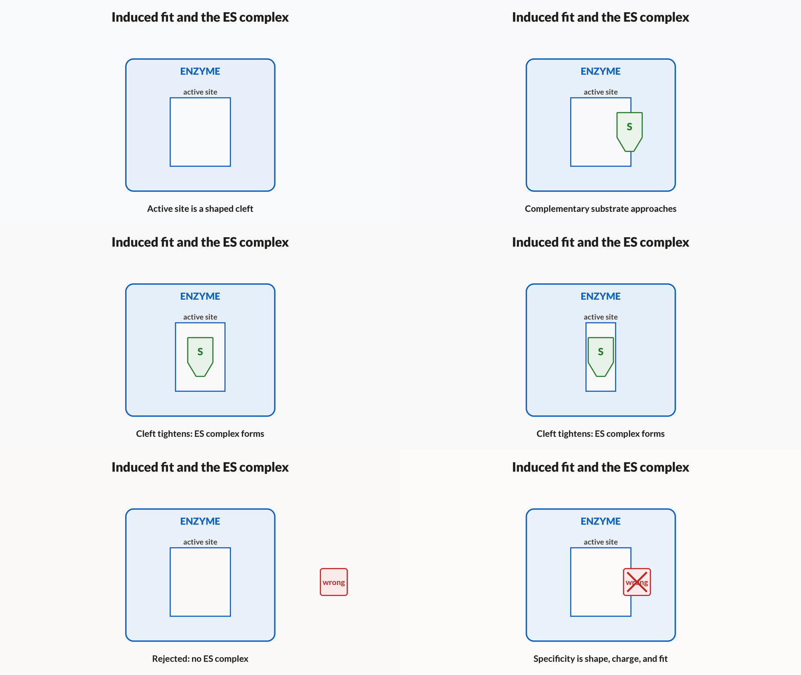
One catalytic cycle
- BindSubstrate diffuses into the active site and is held by weak interactions alone: E + S becomes ES.
- Fit closesThe site adjusts its shape around the substrate, putting the reacting bonds under strain and lining up the catalytic residues.
- ReactThe strained substrate crosses a lowered barrier and is converted while still held: ES becomes EP.
- ReleaseThe product no longer matches the site and leaves; the enzyme, unchanged, takes the next substrate molecule.
Induced fit differs from lock-and-key in that
- The substrate changes shape to match a rigid active site
- The active site changes shape as the substrate binds
- The enzyme will accept any substrate of about the right size
Induced fit moves the enzyme, not the substrate: the site is flexible and closes around what it has caught, and that closing is part of how the barrier is lowered. The first option reverses which partner moves. The third describes no specificity at all — fit is a matter of shape, charge and chemistry together, and size alone would let dozens of wrong molecules in.
6Saturation, and what K_m means
Hold the enzyme fixed and raise the substrate. At low concentrations almost every active site is free, so nearly every substrate molecule that arrives is processed and the rate climbs almost in proportion. As the sites fill, arriving molecules increasingly find them occupied, and the climb flattens. In the limit every site is working every moment: the rate reaches V_max, and no further substrate can improve it.
The whole curve is one equation, V = V_max[S]/(K_m + [S]) — a rectangular hyperbola that approaches V_max but never touches it. K_m is not a rate. It is the substrate concentration at which the enzyme runs at half its maximum, and the lower it is, the less substrate the enzyme needs in order to work near its best.
Figure. The curve is steepest where substrate is scarcest and flattens as the sites fill. The marked point is the definition of K_m: drop from half the V_max line onto the curve and read the concentration underneath. The right-hand end of this plot is at ten times K_m and has still only reached 91% of V_max — the dashed line is approached, never met.
Where K_m comes from
- Start from the rate lawV = V_max[S]/(K_m + [S]), with K_m and V_max fixed for a given enzyme and substrate.
- Set [S] equal to K_mThe right-hand side becomes V_max·K_m/(K_m + K_m), which is V_max·K_m/2K_m.
- CancelV = V_max/2. The half-maximal point is not measured off a graph — it falls out of the equation, and it is the definition of K_m.
| Substrate present | Rate reached | Per cent of V_max |
|---|---|---|
| 0.5 K_m | V_max/3 | 33% |
| K_m | V_max/2 | 50% |
| 2 K_m | 2V_max/3 | 67% |
| 5 K_m | 5V_max/6 | 83% |
| 10 K_m | 10V_max/11 | 91% |
Ninety per cent costs nine times
An enzyme has K_m = 2 mM and V_max = 60 µmol min⁻¹ for its substrate. Find the rate at [S] = 6 mM, and the substrate concentration needed to reach 90% of V_max.
- V = 60 × 6/(2 + 6) = 360/845 µmol min⁻¹
- As a fraction of V_max: 45/600.75
- For 0.9: 0.9(2 + [S]) = [S], so 1.8 = 0.1[S][S] = 18 mM
- 18 mM expressed in units of K_m9 K_m
- V there = 60 × 18/20; the gain over 4554, up only 9
Pro tip. Tripling the substrate from 6 mM to 18 mM buys 9 µmol min⁻¹, having bought 45 by then. Half of V_max costs one K_m; the next 40% of it costs eight more. Read "saturated" in a question as "[S] far above K_m", never as "V has reached V_max".
Two enzymes act on the same substrate. A has K_m = 0.1 mM, B has K_m = 5 mM. Which works better when substrate is scarce?
- A, because it reaches half its maximum rate at a far lower [S]
- B, because the larger K_m means it processes more substrate
- Neither can be judged without knowing their V_max values
K_m is the substrate concentration needed for half-maximal rate, so A is already running at half speed on a fiftieth of the substrate B would need — that is exactly what high affinity means. The second option reads K_m as though it were a rate; it is a concentration, and a big one is bad news. The third confuses two separate questions: V_max says how fast an enzyme runs when saturated, K_m says how much substrate it takes to get there, and scarcity is a K_m question.
7Competitive and non-competitive inhibition
A competitive inhibitor resembles the substrate closely enough to occupy the active site, and does nothing once it is there. Malonate resembles succinate and blocks succinate dehydrogenase this way. Because inhibitor and substrate compete for the same site, whichever is in excess wins: enough substrate restores the rate, so V_max is untouched, and the enzyme merely needs more substrate to reach any given fraction of it — the apparent K_m rises.
A non-competitive inhibitor binds somewhere else on the enzyme and distorts the active site from a distance. The substrate cannot displace it, because they are not competing for the same place. Those enzyme molecules are simply out of service: the top speed falls, while the molecules still working bind substrate exactly as well as before, so V_max drops and K_m does not. Which constant moves is how the two are told apart.
Figure. The two marked points sit at exactly the same height — half of V_max — and differ only in how far right they lie: here the inhibitor has tripled the substrate needed to get there. Both curves climb towards the same horizontal V_max line, which is what "beatable by excess substrate" looks like on a graph. A non-competitive inhibitor would look entirely different: the horizontal line itself would drop, and the half-maximal point would not move sideways at all.
| Feature | Competitive | Non-competitive |
|---|---|---|
| Where it binds | the active site itself | a separate site on the enzyme |
| Resembles the substrate | yes — that is why it fits | no |
| Apparent K_m | raised | unchanged |
| V_max | unchanged | lowered |
| Does more substrate help? | yes, it out-competes the inhibitor | no, the blocked enzyme cannot be freed by substrate |
Buying the rate back with substrate
The enzyme of the previous concept (K_m = 2 mM, V_max = 60 µmol min⁻¹) is run at [S] = 6 mM, where it managed 45 µmol min⁻¹. A competitive inhibitor is added at [I] = 4 mM; its inhibition constant is given as K_i = 2 mM. Find the new rate, and the substrate concentration that would restore 45 µmol min⁻¹.
- Apparent K_m = K_m(1 + [I]/K_i) = 2(1 + 4/2)6 mM
- V = 60 × 6/(6 + 6) = 360/1230 µmol min⁻¹
- Fraction of the uninhibited rate: 30/45two thirds
- For V = 45 again: 45(6 + [S]) = 60[S], so 270 = 15[S][S] = 18 mM
- Uninhibited enzyme at that same 18 mM54 µmol min⁻¹
Pro tip. Tripling the substrate restores the rate the enzyme had before the inhibitor arrived — but the uninhibited enzyme would now be doing 54, so the inhibited curve never catches the free one at any finite [S]. "Overcome by excess substrate" means the two curves share the same V_max in the limit, not that the inhibition disappears.
An inhibitor lowers an enzyme's V_max but leaves its K_m unchanged. It is
- Competitive, and can be beaten with excess substrate
- Non-competitive, binding away from the active site
- An activator working at an allosteric site
Capping V_max is the signature of an inhibitor that is not competing for the active site: it binds elsewhere, takes those enzyme molecules out of service, and no amount of substrate can displace it. A competitive inhibitor is the mirror image — it raises the apparent K_m and leaves V_max alone, precisely because excess substrate does win the site back, so the first option has both constants wrong. An activator would raise the rate rather than lower it.
8Optimum temperature and optimum pH
Warming a reaction speeds it up: molecules collide more often and more of them arrive with enough energy to react. For a human enzyme this holds until about 37 °C. Past that, a second process takes over — the weak bonds holding the tertiary fold begin to give way, the active site loses its shape, and the number of working enzyme molecules collapses. Above about 45 °C the collapse dominates completely.
So the temperature curve is not a symmetric bell. It rises gently over tens of degrees and falls off a cliff over a few, and the two halves mean different things: cooling an enzyme merely slows it and is fully reversed by rewarming, whereas heating past the optimum denatures it and is generally not reversed by cooling. pH acts differently again. It changes the ionisation of the acidic and basic side chains that build and hold the active site, so each enzyme has an optimum matching the compartment it works in.
Figure. Look at the two sides of the peak, not the peak itself. The climb takes some thirty degrees; the fall is essentially over within ten. That asymmetry is the whole point — the rise is ordinary chemistry speeding up, the fall is the protein being destroyed — and it is why drawing this as a tidy symmetric bell teaches the wrong biology. The left half is reversible, the right half is not.
| Enzyme | Optimum pH | Where it acts |
|---|---|---|
| Pepsin | about 2 | stomach, in hydrochloric acid |
| Salivary amylase | about 6.8 | mouth, in near-neutral saliva |
| Trypsin | about 8 | small intestine, in alkaline pancreatic juice |
An enzyme has almost no activity at 0 °C and almost none at 60 °C. Warming the cold sample to 37 °C restores it fully; cooling the hot one to 37 °C restores nothing. Why?
- Cold destroys the active site, while heat only slows the molecules
- Cold only slows the reaction; heat unfolds the protein
- Both are denatured, but the cold sample refolds more easily
Below the optimum the protein is entirely intact and the loss is kinetic — fewer collisions, less energy in each — so warming recovers all of it. Above roughly 45 °C the weak interactions holding the fold give way, the active site loses the shape that made it a site at all, and that change is generally irreversible, so cooling recovers none of it. The first option has the two halves swapped, and the third would predict at least partial recovery in the hot sample, which is not what is seen.
Notes
- The main biomacromolecules are carbohydrates (energy and structure), proteins (chains of amino acids), lipids (energy storage and membranes) and nucleic acids (DNA and RNA).
- Proteins: 20 amino acids link by peptide bonds, with primary (sequence), secondary (helix/sheet), tertiary (3-D fold) and quaternary (multiple subunits) structure.
- Enzymes are biological catalysts (mostly proteins) that lower activation energy, are highly specific (lock-and-key or induced-fit) and are reusable.
- Enzyme activity depends on temperature, pH and substrate concentration, and each enzyme has an optimum pH and temperature.
- Cofactors and inhibitors: coenzymes and metal-ion cofactors aid catalysis; a competitive inhibitor resembles the substrate (e.g. malonate inhibits succinate dehydrogenase).
Formulas
- Peptide bond links the –COOH of one amino acid to the –NH₂ of the next
- Enzymes act by lowering activation energy (E_a)
- Michaelis-Menten: V=\frac{V_{max}[S]}{K_m+[S]}
- Competitive inhibition raises K_m while V_{max} is unchanged
- Primary structure (amino acid sequence) determines all higher structures
Exam traps & shortcuts
- Enzyme names usually end in '-ase' and indicate the substrate or reaction (lactase, dehydrogenase).
- A competitive inhibitor resembles the substrate and can be overcome by raising substrate concentration.
- Above the optimum temperature, enzymes denature (lose tertiary structure) and irreversibly lose activity.
Reference tables
Every line here should be reconstructible from the concept it came from, not merely recalled.
| Quantity | Relation | Watch for |
|---|---|---|
| Peptide bonds | n residues in one chain give n − 1 bonds | one water out per bond; count per chain, not per protein |
| Chain mass | roughly 110 Da per residue | an average, and it already excludes the lost water |
| Rate law | V = V_max[S]/(K_m + [S]) | a hyperbola: V_max is approached, never reached |
| Meaning of K_m | [S] = K_m gives V = V_max/2 | a concentration, not a rate; low K_m is good at low [S] |
| Competitive inhibitor | apparent K_m = K_m(1 + [I]/K_i), V_max unchanged | excess substrate restores the rate |
| Non-competitive inhibitor | V_max falls, K_m unchanged | excess substrate does not help |
| Catalysis | lowers E_a, equally in both directions | the free-energy change and the equilibrium are untouched |
| Structure | primary sequence fixes every higher level | denaturation destroys levels 2 to 4 and spares level 1 |
Apoenzyme (protein alone) plus cofactor gives holoenzyme (the working catalyst). Strip the cofactor away and the apoenzyme cannot catalyse anything.
| Kind of cofactor | How it is held | Example |
|---|---|---|
| Prosthetic group | tightly bound and permanently resident | haem in catalase and peroxidase |
| Coenzyme | associates only transiently, during the reaction | NAD and NADP, built on the vitamin niacin |
| Metal ion | coordinated within the active site | zinc in carboxypeptidase |
Recap
Read only this the night before.
- Families
- Sugar–glycosidic, amino acid–peptide, nucleotide–phosphodiester. A fat is glycerol plus three fatty acids and repeats nothing, so it is not a true polymer.
- Counting
- n residues in one chain give n − 1 peptide bonds and n − 1 waters. Two chains means doing it twice.
- Levels
- Primary is covalent and survives boiling. The active site is a tertiary shape and does not.
- Catalysis
- Lowers the barrier in both directions. Rate changes, equilibrium and yield do not.
- Saturation
- [S] = K_m gives exactly half of V_max; even ten times K_m reaches only 91%.
- Inhibition
- Competitive moves K_m and is beaten by substrate. Non-competitive moves V_max and is not.
- Optima
- Cold slows and is reversible; heat above the optimum denatures and is not. The temperature curve is lopsided.
Practise Biomolecules and Enzymes
Reading is free and needs no account. Practice, mocks and progress live in the app.
- 5 exam-style questions on this topic, with explanations
- A 5-question practice set that ends the chapter
- Timed mocks scored with the real marking scheme
- Readiness tracked per topic, kept on your device